Single-Celled Stentors - The Giants In A Microscopic World

The microscopic world of unicellular organisms is a large one. Hidden beyond our eyesight lie countless of the little creatures that jerk around sporadically, feeding on bits of nutrients in the air or otherwise engulfing each other up. This behaviour happens regularly; if you ever take a good optical microscope and use its highest magnifications to see them, you might find entire communities of species constantly interacting with each other in a bizarre cacophony of life. Such events might involve bacteria, unicellular fungi (e.g. yeast) and even microalgae.
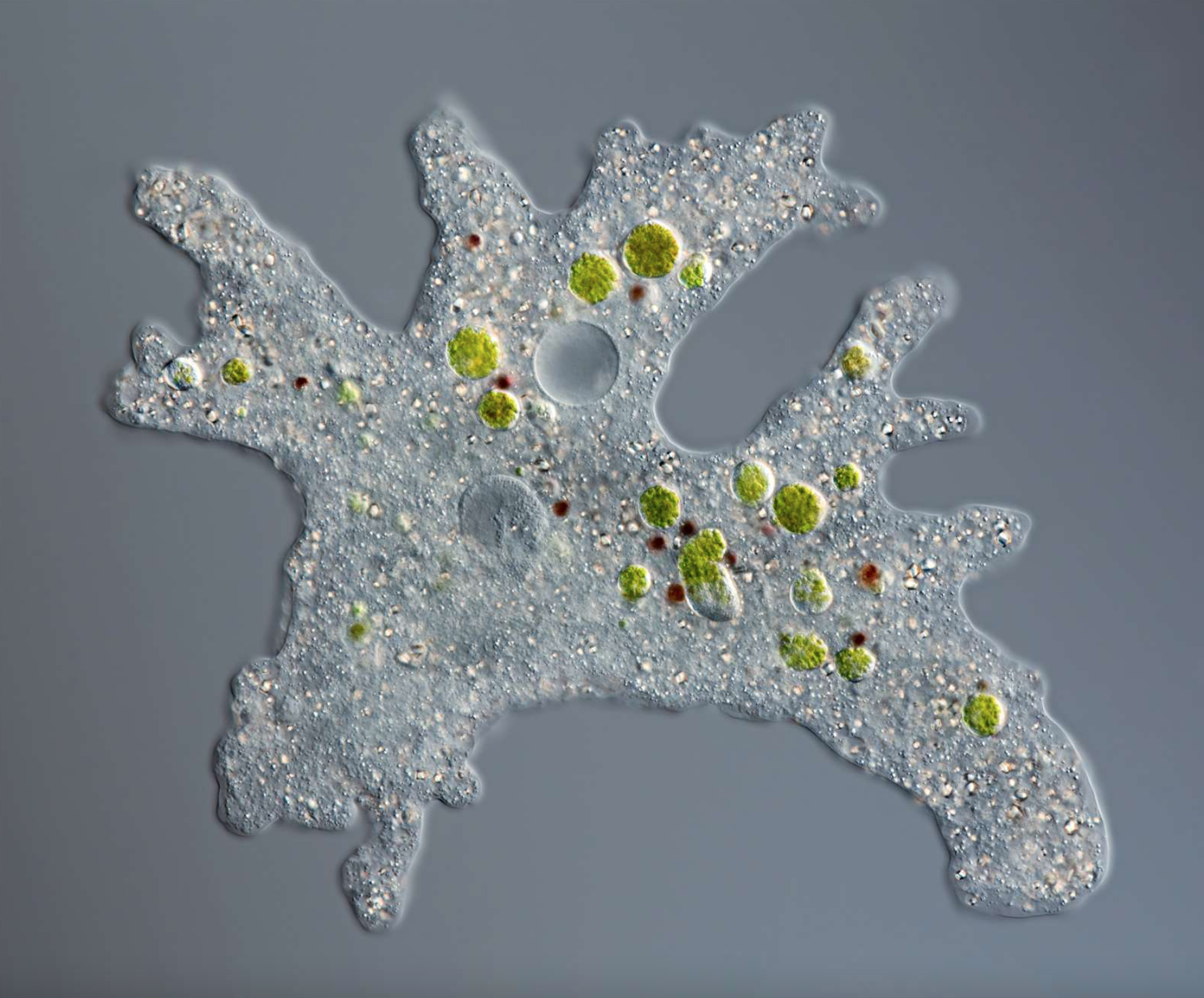
In between those critters, you may also find what would look like giants under the lens. Stomping and blobbing their way across their microenvironment are the amoebas; amorphous protists that stretch out tentacle-like pseudopodia to wrap themselves around and engulf smaller microbes. These appear everywhere, and could be seen almost like apex predators in their little world. In addition to these, creatures that suck up nearby particles like vaccuums roam, and they are known as filter feeders. Such organisms can be compared to decomposers, which feed on the organic wastes of other organisms, for their passive way of obtaining their food.
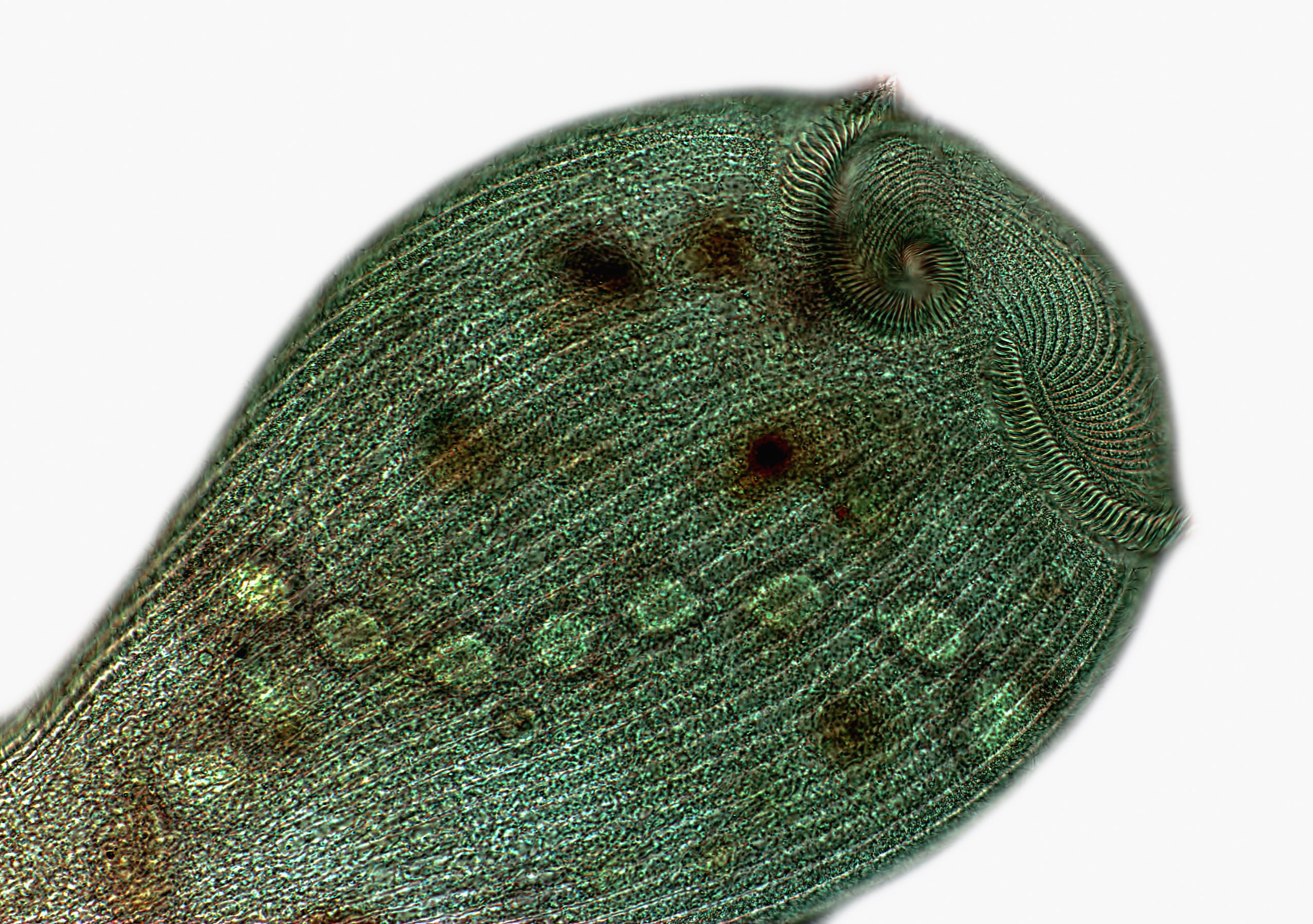
One particularly significant filter feeder, studied numerous times for its nigh-on universal presence around the world, is the stentor. Measuring up to 4 millimetres in length, it is one of the largest unicellular organisms in the planet and in clear water can even be visible to the naked eye. If you do happen to witness one, you will find that stentors look somewhat like trumpet horns, with a large mouth at one end thinning off into a pointed section at the other end. This unusual shape and their scavenging mannerisms once made scientists refer to them as trumpet animalcules - which is silly. You see, the term 'animalcules' is really just a fancy name for microscopic animals, and we now know that stentors are not animals at all! Instead, they are protozoans, or animal-like protists. (If you wish to know more about them, protists as a group are covered more thoroughly in my seaweed post.)
This time I will be focusing on this particular protist - mostly because I find them interesting, but also as a way to give the suckers some spotlight before the public. They go unnoticed too often, I think, what with all the talk on bacteria and yeast cells. So, let's take a closer look at their insides, shall we?
Filter Feeding & Swimming With Jetpacks
The better the quality of our microscope, the more clearly we can view the the going-ins and outs of a stentor. As such, let's assume that our microscope costed our scientific institution some ten thousand dollars (all financed by them, of course) and is maintained under the highest of quality controls possible. For real, though, who wouldn't want to have one of these for their birthday?
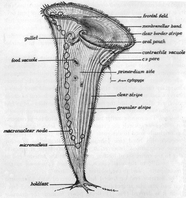
Using our mega-microscope, we find a ring of tiny hairs covering the entirety of the stentor's mouth and directed towards its throat/gullet. These are called cilia, and they serve to easen the process of ingesting food by catching microbes like bacteria in the water and directing them inside the stentor. They also vibrate and move in circular patterns, creating small vorteces that pull water (which may contain even more little bits of food) inside, creating the 'vaccuum' effect that I spoke of earlier, with other cilia around its body helping it swim and move around. This, I should note, is also why you may hear stentors be referred to as ciliates - because they have cilia. You may further liken this to the cilia present on the epithelial cells that line your own trachea (i.e. the main respiratory tract), which serve a similar purpose in moving particles through the throat.
Once inside the gullet, food is processed and filtered. Here, any desirable material is absorbed for future use, with the undesirable stuff (like inorganic material or toxins) being expelled - hence its name of filter feeding.
Additionally, stentors are apparently so large that they not only feed on organic particles in the water, but have been known to consume even multicellular organisms like water fleas! Though it is astonishing to me, it honestly sounds like a nightmare for the poor flea. Anyway.
In order to feed peacefully and consistently, stentors use their tail-piece of their thinner end to attach themselves to a preferred substrate. This piece is known as the holdfast, and it can anchor the cell down even with its large body. Of course, this will not work in turbulent or convecting waters, which is why stentors generally live in more placid, stagnant bodies of water. Since they also tend to inhabit freshwater environments, the salinity inside their bodies will in most cases be higher than their surroundings, prompting water to constantly flow into them via osmosis (which is the movement of water through a semi-permeable membrane from a high solute concentration to a low one). Accordingly, stentors have evolved ways to handle their sudden influx of water - namely, they push it out. Literally.
Inside the stentor resides a contractile vacuole. This organelle is used to store water, and it is surrounded by a thin membrane that allows water to leave or enter freely - so long as it remains relaxed. Now, a number of other organisms also have their own types of water-storing vacuoles (such as plant vacuoles, which are made to store the sugary water that you might know as 'sap'), but contractile vacuoles are particularly unique. These are operated by contractile structures in the cell - the microtubules and myonemes that make up its cytoskeleton. In principle, microtubule fibers are long, fibrous proteins, while myonemes are formations of filaments placed in rows - but, though they have different pathways that allow them to contract and relax, both are influenced by the positively charged ions in the stentor. Additionally, they can be distinguished with antagonistic functions, so that when one increases in length, the other shortens (e.g. calcium ions make the myonemes contract and microtubules relax). When the contractile structures attached to the vacuole contract/relax, the vacuole can shrink rapidly to expel excess water outside. These jets of water can then be used by the stentor to propel itself more quickly across the water (and yes, this does mean that stentors have in-built jetpacks).
Interestingly, though stentors are unicellular, they have been found to display other functions that are uncannily similar to multicellular organisms' - including self-regeneration and the presence of multiple nuclei. And so we dig deeper.
Divided Control & Regeneration
In case you don't know, most eukaryotic cells - whether they exist as individual organisms or form part of a bigger multicellular creature - contain only one nucleus. This is typically the largest organelle in the cell, and it is the main characteristic that defines eukaryotes (with prokaryotes having no nucleus). Its principal functions are to store genetic information in the form of DNA supercoiled around proteins, and to control the cell's metabolism by directing that genetic information to move around the cell and give orders to its machinery. Such is the case for all eukaryotic cells - but there are often differences in their methodologies.
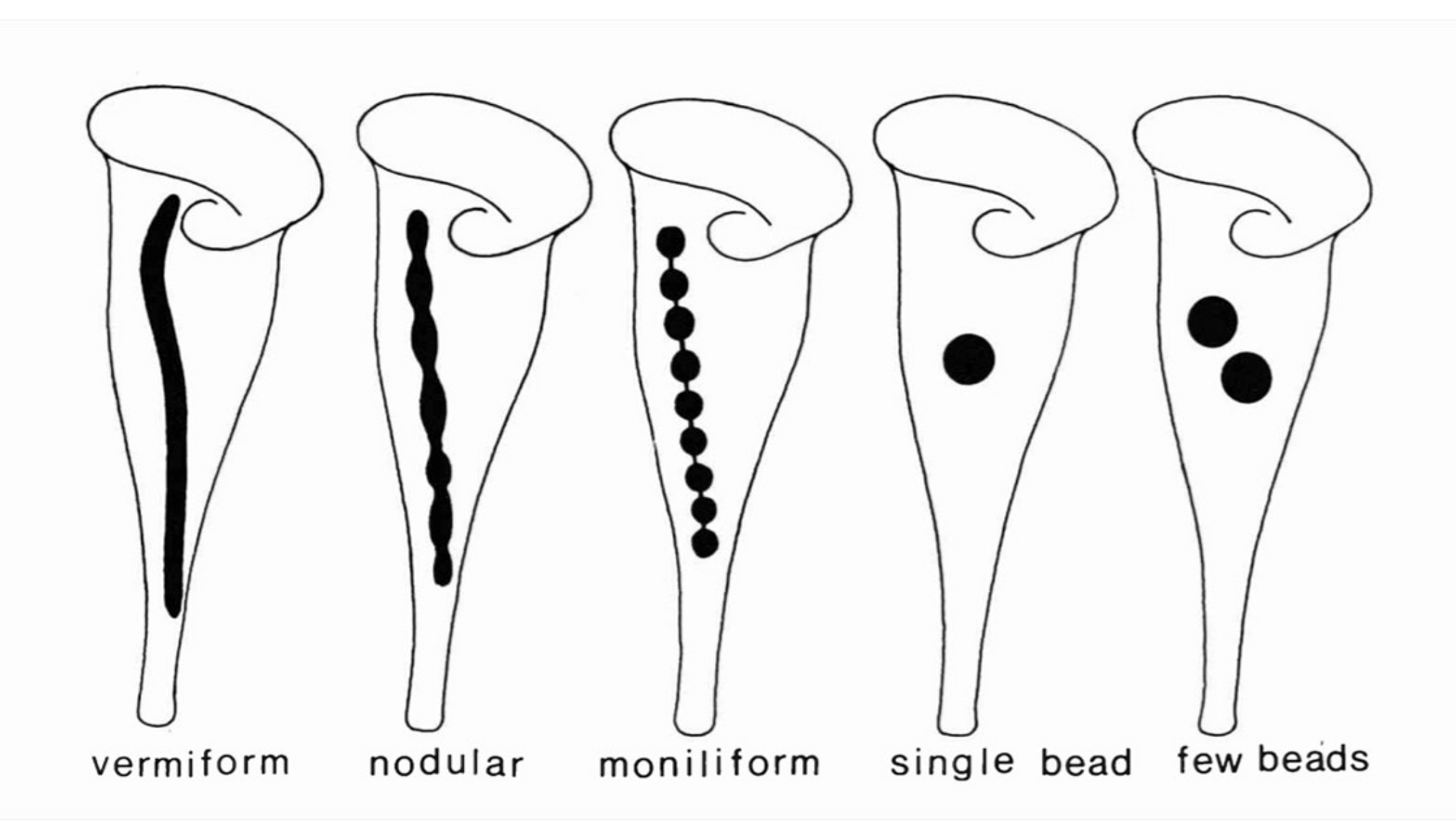
In the case of the stentor, its enormous size makes it difficult for a single nucleus to control all the chemical processes in its body. One nucleus simply wouldn't be able to reach. As such, stentors tend to split the work between multiple centers of control: first with a number of small micronuclei, which govern how a stentor divides by a very specific type of sexual reproduction (known as conjugation); and then with a singular large macronucleus, which is in charge of all the asexual processes in the cell, from feeding and sensing to protein synthesis. This division of control is another characterising feature in ciliates, but the stentor takes it yet another step further. Instead of its macronucleus being a large blob with a defined area, it can have an elongated, moniliform shape that appears similar to a string of beads (which are referred to as nodes). Depending on the species of stentor, this may also be vermiform (like a thick rope), nodular (like a very strange worm), a spherical blob or just a few isolated blobs. Why this is the case, nobody knows (yet), but it is nonetheless a good way to distinguish one stentor species from another.
For instance, the species Stentor araucanus, first observed in South African lakes, is vermiform and living a wonderfully unique life amongst its peers. Having multiple nuclei further allows stentors to exhibit astounding levels of regeneration; if you somehow manage to cut one in two, for example, its two halves will eventually become their own independent organisms with fully functional metabolisms. How this occurs is beyond this post, but I'm sure it would be a fascinating topic to investigate!
So, now that you know a little more about the microscopic world around you, what do you think of it? If it did not make much of an impact, worry not; the stentor is only one in a million of interesting case studies to explore and digest. Quite a lot for small things!
References
- Ward, B. (n.d.). Stentor: Structure, Classification, and Characteristics. Microscope Clarity. Retrieved from https://microscopeclarity.com/stentor/
- Edmonds, M. (2021). What Is Filter Feeding? HowStuffWorks. Retrieved from https://animals.howstuffworks.com/animal-facts/filter-feeding.htm
- Huang, B. & Pitelka, D. R. (1973). The contractile process in the ciliate, Stentor coeruleus. I. The role of microtubules and filaments. Journal of Cell Biology. Retrieved from https://pubmed.ncbi.nlm.nih.gov/4633444/
- Thamm, M. et al (2010). Insights Into the Phylogeny of the Genus Stentor (Heterotrichea, Ciliophora) with Special Emphasis on the Evolution of the Macronucleus Based on SSU rDNA Data. Acta protozoologica. Retrieved from https://www.ejournals.eu/Acta-Protozoologica/2010/Issue-3/art/3716